
Melbourne knee surgeon Parminder J Singh treats knee pain for some conditions using arthroscopic (keyhole) knee surgery. The most common conditions comprise meniscal tear and anterior cruciate ligament (ACL) tears. The rest of this article will focus on ACL injuries.
Anatomy of the ACL
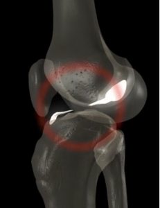 The ACL is one of 2 cruciate ligaments which aids in stabilization of the knee joint. It is a strong band made up of tissue (connective tissue and collagenous fibers) that originates from the shin bone (anteromedial aspect of the intercondylar region of the tibial plateau) and extends into the thigh bone (postero-medially to attach to the lateral femoral condyle).
The ACL and the posterior cruciate ligament (PCL) together form a cross within the knee joint and prevents excessive forward or backward motion of the shin bone (tibia) in relation to the thigh bone (femur) during bending (flexion) and straightening (extension).
The ACL additionally provides rotational stability to the knee with varus (force from inside to outside) or valgus (outside to inside) stress.
 What is the mechanism of ACL injury?
Most ACL tears occur in athletes by non-contact mechanisms and contact mechanisms, such as:
- Rotational forces versus a direct hit to the knee.
- Sudden changes in the direction of movement, rapid stopping, jumping and landing abnormally,
- A direct blow to the outer aspect of the knee, or slowing down while running.
Who is at risk of an ACL injury?
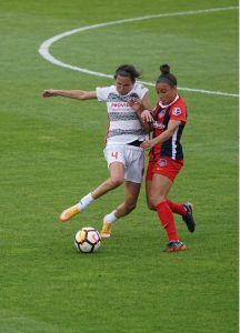 The high-risk athletes for non-contact injury include skiers, soccer players, and basketball players, while the most at risk athletes for contact injury are football players.
Some studies suggest that females may have weaker hamstrings and preferential utilize the quadriceps muscle group while decelerating. When engaging the quadriceps musculature while slowing down, this places abnormally increased stress on the ACL. (the quadriceps muscles are less effective at preventing anterior tibial translation versus the hamstring muscles). A second factor that may increase the risk of ACL injury is the increased valgus angulation of the knee when changing direction.
Assessment of an ACL injury
The majority of individuals when they injure their ACL report hearing and or feeling a sudden pop. This is often associated with a sudden onset of swelling and instability of the knee.
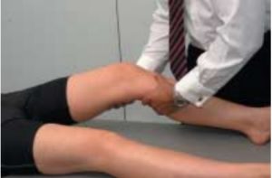 Assessment of the knee usually comprises inspection of the joint for any swelling or deformity. Palpation of the knee may reveal tenderness of the inside of the knee (medial side) and the joint line indicating associated injuries of the knee such as medial collateral knee injury (MCL) and meniscal tears. Moving the knee may reveal more information regarding the ligaments.
- The test to assess the ACL integrity is called the anterior draw test (sensitivity of 92% and specificity of 91% in chronic injuries, but not acute injuries.)
- Lachman’s test (This test has a sensitivity of 95% and specificity of 94% for ACL rupture)
- Pivot shift test (highly specific test (98%)) when positive. A comparison with the other knee usually reveals laxity in the affected knee in the presence of an ACL injury.
Diagnosis
Magnetic resonance imaging (MRI) scan of the knee is often used to confirm the diagnosis of an ACL tear. MRI scans have a sensitivity of 86% and specificity of 95%. Secondary signs on the MRI scan may also depend on the severity of the initial injury and include bone marrow swelling fracture and associated medial collateral ligament injury. Diagnosis may also be made with arthroscopic knee surgery to differentiate complete from partial tears
 Management of an ACL injury
Following the initial injury, REST, ICE, COMPRESION, ELEVATION of the affected knee should be considered. Initially, the patient may need crutches and may be non – weight bearing. Pain relief should also be considered.
ACL injuries should be referred to an orthopaedic surgeons to discuss the options of treatment comprising non-surgical treatment or arthroscopic knee surgery with a view to reconstruction of the ACL and treatment of any associated injuries.